Earn CME credits for Lab Re-Accreditation
These modules are Self-Study activities designed for physicians to maintain credentials as well as update their interpretive and didactive skills. The materials will be presented in an on-line format where the student will evaluate didactic material, images and animations relevant to the study of Nuclear Cardiology.
Each course topic concludes with an interactive quiz to guage the learner's retention of the materials presented.
Disclosures: All Faculty, CME Planning Committee Members, and the CME Office Reviewers have disclosed that they do not have any relevant financial relationships with commercial interests that would constitute a conflict of interest concerning this CME activity.
Additional Course Information
Comprehensive review of Patient preparation, Orbit, Matrix and Gating.
Discuss Reconstruction, Reorientation, Segmentation, Sampling, Polar Maps & Quantitative Perfusion.
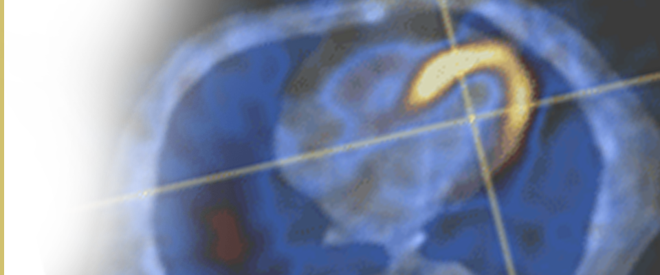
Patient Related Artifacts, Errors and Pitfalls
Improve identification and handling of Errors and Artifacts.
Identify motion artifacts, regional & diffuse soft tissue attenuation and reconstruction artifacts.
Proper usage of motion and attenuation correction.
Gamma Camera and Quality Assurance Testing
Explain how a gamma camera works and how SPECT images are formed in relation to collimation, crystal technology, photomultiplier tubes and essential electronic components.
The application of solid state detectors in nuclear cardiology.
Evaluate NEMA recommendations and identify artifacts in nuclear cardiology quality control.
Image Interpretation
Identify proper image interpretation and artifact in rotating planar imaging.
Interpret standard nomenclature and slice orientation in SPECT imaging.
Analysis of volumes & ejection fraction and identifying wall motion & thickening in Gated SPECT imaging.
Quantitative and Qualitative assessments in image interpretation.
Radiopharmaceuticals & Radiation Safety
Applying the characteristics of individual tracers, concepts of tracer kinetics, extraction, distribution, retention and radiation physics of radiopharmaceuticals in nuclear cardiology.
Addressing radiation safety concerns in nuclear cardiology in regard to: terminology, time, distance, shielding, biological effects, monitoring, occupational exposure, stochastic effects and deterministic effects.
ALARA dose considerations.
Stress and Imaging Protocols
Compare and contrast exercise and pharmacologic stress testing options in terms of application, accuracy, strengths and weaknesses.
Imaging protocols in Nuclear Cardiology under administration of Thallium and TC-99m.
Apply principles of nuclear cardiology to board exam preparation.
Dose Reduction in Nuclear Medicine and PET/CT
Appreciate the Magnitude of the Issue/Epidemiology of Radiation in Medical Imaging. Apply principles of dose versus scan time, radiation units, radiation from nuclear medicine studies. Differentiate types of ionizing radiation, biological, physical, and effective half-life and an overall general approach to managing dose. Recognize crystal technology/camera design, reconstruction algorithms, new radiotracers and Imaging protocols in nuclear medicine. Evaluate special considerations with pediatric patients, modified dose chart and Image quality. Identify future directions regarding the co-evolution of camera technology, software, and hardware as well as a multi-faceted approach to dose reduction.
Generator Safety in Nuclear Medicine
Comprehensive background in Mo/Tc and Sr/Rb generators as well as functionality and special considerations. Recognize the rationale and implication surrounding the FDA recall of the Rb-82 generator for PET myocardial perfusion imaging. Discuss the Rb-82 generators as a paradigm for the principles in generator safety.
Positron Emission Tomography (PET)
Understand the basic principles of PET radiation physics
Compare SPECT vs PET
Review the components of a PET camera
Understand the workflow for image processing and reconstruction
Review the available radiotracers for perfusion and viability
Discuss Stress Protocols
Multigated Acquisition (MUGA)
Review Techniques for Evaluating Left Ventricular Function
Discuss Planar and SPECT ECG gated imaging
Understand the workflow for FRNA and first Pass
Review Techniques for Evaluation of Right Ventricle function
Discuss First Pass and SPECT ERNA
Review the evaluation of Left to Right Shunt
MD Training faculty believe that in this ever changing medical environment where imaging is heavily scrutinized and ever evolving, continued education as well as board certification and recertification in Nuclear cardiology will become ever more important to assure competence, cutting edge practice, reimbursement and credentialing. Guideline based medicine practice is clearly the trend in this and all fields especially when prescribing diagnostic radiation. Ever awareness of radiation exposure is mandatory. This course’s content covers these techniques and protocols, which are ever evolving, and outlines their applications to the participant.
Director of Clinical Education
MD Training @home, LLC
Board certified in Internal Medicine and Cardiology
Subspecialty board certification: Echocardiography, Nuclear Cardiology, Vascular Imaging
The University of Arizona College of Medicine – Tucson designates this enduring materials activity for a maximum of 20 AMA PRA Category 1 Credit(s)TM. Physicians should claim only the credit commensurate with the extent of their participation in the activity.
Current CME Approval Period: 5/26/2023 to 5/25/2025
Date of Last Review: 5/26/2023
Original Release Date: 8/09/2009
UA Continuing Medical Education
PO Box 245121
Tucson, AZ, 85724-5121
(520)626-7832
FAX (520)626-2427
uofacme@u.arizona.edu
www.ocme.arizona.edu